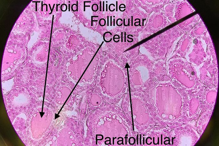
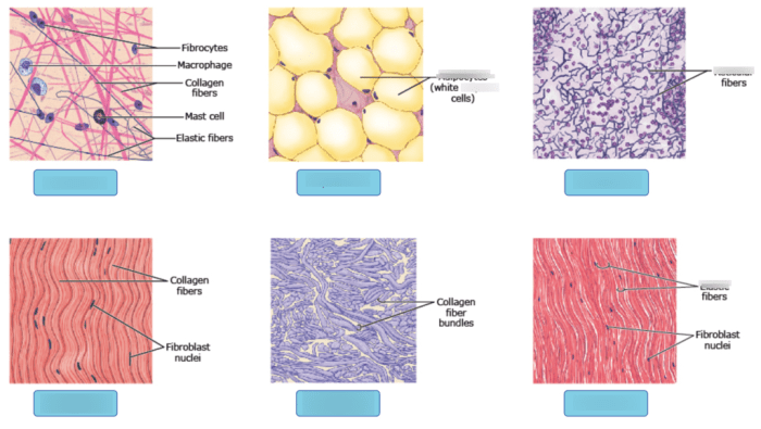
As we embark on the intricate exploration of label the tissue and structures on this histology slide, we delve into a realm where meticulous observation and precise terminology converge to unveil the secrets of human biology. This comprehensive guide will navigate you through the fascinating world of tissue identification, structure labeling, and histological techniques, empowering you with the knowledge to decipher the complexities of the human body.
Through the lens of this histology slide, we will uncover the diverse tapestry of tissues, each with its unique characteristics and functions. We will meticulously label the major structures, unraveling their intricate relationships and significance within the tissue architecture. This journey will not only enhance your understanding of human anatomy but also equip you with the skills to interpret histological slides, a crucial tool in medical diagnosis and research.
Tissue Identification

The histology slide reveals a variety of tissues, each with distinct characteristics and functions. These tissues can be classified into four main types: epithelial, connective, muscle, and nervous.
Epithelial Tissue
- Consists of closely packed cells that form a protective barrier
- Examples: Skin, lining of organs, glands
Connective Tissue
- Provides support, protection, and nourishment to other tissues
- Examples: Bone, cartilage, adipose tissue
Muscle Tissue
- Responsible for movement
- Examples: Skeletal muscle, smooth muscle, cardiac muscle
Nervous Tissue
- Transmits electrical signals to control body functions
- Examples: Neurons, glial cells
Structure Labeling
The histology slide showcases several key structures, each playing a vital role in the overall function of the tissue.
Epithelial Layer, Label the tissue and structures on this histology slide
A layer of epithelial cells that forms the outermost layer of the tissue
Basement Membrane
A thin layer of connective tissue that separates the epithelial layer from the underlying connective tissue
Connective Tissue Layer
A layer of connective tissue that provides support and nourishment to the epithelial layer
Blood Vessels
Thin-walled tubes that transport blood to and from the tissue
Tissue Architecture

The tissue in the histology slide exhibits a well-organized architecture that contributes to its overall function.
The epithelial layer forms a protective barrier against external factors, while the underlying connective tissue provides support and nourishment. The blood vessels ensure a continuous supply of nutrients and oxygen to the tissue.
Histological Techniques: Label The Tissue And Structures On This Histology Slide
The histology slide was prepared using a series of histological techniques that allow for the visualization and study of the tissue.
These techniques include:
- Fixation: Preserving the tissue’s structure
- Embedding: Enclosing the tissue in a solid medium for slicing
- Sectioning: Cutting thin slices of the tissue
- Staining: Adding dyes to enhance the visibility of specific structures
Clinical Relevance

The histology slide can be used for diagnostic and monitoring purposes in various clinical settings.
By examining the tissue’s structure and identifying any abnormalities, healthcare professionals can diagnose diseases, assess their severity, and monitor treatment response.
For example, a biopsy of skin tissue can be used to diagnose skin cancer, while a biopsy of lymph node tissue can help detect the presence of metastatic cancer cells.
Top FAQs
What is the purpose of label the tissue and structures on this histology slide?
Labeling the tissue and structures on a histology slide allows for the accurate identification and characterization of various tissues and their components. This information is crucial for understanding the normal structure and function of tissues, as well as for diagnosing and monitoring diseases.
How are tissues identified on a histology slide?
Tissues are identified based on their unique microscopic characteristics, including cell morphology, tissue architecture, and staining patterns. Histologists use specific stains and dyes to highlight different tissue components, making it easier to distinguish between different cell types and structures.
What are the different types of structures that can be labeled on a histology slide?
The types of structures that can be labeled on a histology slide vary depending on the tissue being examined. Common structures include cells, nuclei, cytoplasm, extracellular matrix, blood vessels, nerves, and glands.
How are histological techniques used to prepare a histology slide?
Histological techniques involve a series of steps to prepare a tissue sample for microscopic examination. These steps include tissue fixation, embedding, sectioning, and staining. Each step is carefully controlled to preserve the tissue’s structure and allow for optimal visualization under a microscope.
What is the clinical relevance of label the tissue and structures on this histology slide?
Labeling the tissue and structures on a histology slide is essential for medical diagnosis and research. By accurately identifying and characterizing tissues, pathologists can diagnose diseases such as cancer, infections, and autoimmune disorders. Histology slides are also used to monitor disease progression and treatment response.